Key Areas of Research
Areas of Research
Biomineralization & Hard Tissue Regeneration
Dentin, enamel and bone are highly mineralized hard tissues, uniquely comprised of carbonated hydroxyapatite nanocrystal and organic components in a controlled manner which confers them with elaborate morphologies and outstanding mechanical properties. Our research focuses on understanding the interplay of organic components and the mineral in bio-mineralization process. Current researches include:
- Biomimetic mineralization of collagen fibrils using the polymer-induced liquid-precursor process, where anionic polymers are used to induce intrafibrillar mineralization in collagen fibrils. This project attempts to elucidate the role of acidic non-collagenous proteins (NCPs) in stabilizing calcium phosphate ions in solution but nucleating crystallization in organic matrix.
- Mimicking nanostructure of bone and dentin using elastin-like recombinant polypeptides as an analogue of collagen. Elastin-like polypeptides can self –assemble into a hierarchical organization at multiple length scales ranging from an ordered and dynamic β-spirals to 5-nm diameter filaments and to fibrils with a diameter less than a few hundred nanometers. Because their superstructure and physicochemical properties can be fine-tuned, mineralization of elastin-like recombinant polypeptides might help understanding the role of electrostatic interactions and capillary forces in biomineralization.
- Remineralization of Enamel Lesions. Dental caries continues to be a major health issue in both adults and children. We attempt to develop remineralization methods that can be used to repair tooth decay at its early signs.
Biomedical Surfaces & Interfaces
The engineered biomodification of surfaces and interfaces for dental and other biomedical applications is one of the main topics in biomaterials science and engineering. At the MDRCBB we have focused on a) understanding basic bio/non-bio interactions [Biointerphases, (2012)7, pp48 and Acta Biomaterialia, (2010) 6, p291 ] and 2) using oligopeptides and recombinant biopolymers to coat biosurfaces and natural tissues to improve performance of dental and orthopedic materials and devices [Book-Biological and Mecial Physics-Biomedical Engineering Series, (2013) pp105]. We have been able to design and produce antimicrobial [Acta Biomaterialia, (2013)9, pp8224], biomineralizing [Advanced Healthcare Materials (2014) 9, pp8224], hydrophobic [PLoS ONE, (2014)9, 311579], bioactivating [Colloids and Surfaces B: Biointerfaces, (2014)114, pp225], and multifunctional surfaces [Colloids and Surfaces B: Biointerfaces, (2013)107, pp189]. Some examples of the medical products that will be benefited from the development of these technologies are dental implants, esthetic restorations, orthopedic implants, and orthodontic appliances.
Characterization of Dental Materials
Polymerization & Shrinkage
Polymerization shrinkage of dental resin composites causes premature failure of composite restorations through interfacial debonding. The MDRCBB has developed an innovative suite of nondestructive techniques and analytical methods for characterizing polymerization shrinkage and the damage it can cause to composite-restored teeth. These include digital image correlation to capture full-field shrinkage strain in real time [Dental Materials, (2015) 31, pp391], acoustic emission measurement to detect and monitor interfacial debonding [Dental Materials, (2016) 32, pp742], and micro-CT to quantify interfacial leakage in 3D [Dental Materials, (2015) 31, pp382]. A constitutive model has also been developed to derive the temporal changes in the mechanical properties of resin composites and to predict the level of shrinkage stress in a composite restoration [Dental Materials, (2013) 29, pp1108].
Simulation of Oral Challenges
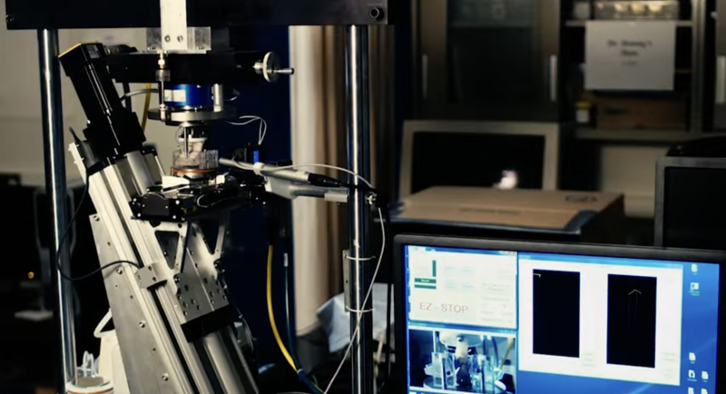
Artificial Resynthesis Technology (ART)
Dental materials scientists are constantly searching for new restorative materials and they want to know how the new materials perform clinically. Testing the new materials in human volunteers would require safety approval and will take considerable time. A groundbreaking piece of research leading to the formation of the MDRCBB had been the development of the world’s first chewing machine here in Minnesota [IEEE Transactions on Biomedical Engineering, (1991) 38, pp339]. As the name indicates, ART resynthesizes or replicates that part of the chewing cycle in which the teeth come into contact. The MDRCBB’s chewing machines can perform both 2- and 3-body wear tests, completing the annual equivalence of 300,000 cycles of chewing in less than a day [Dental Materials, (1985) 1, pp238].
Oral Hygiene Simulator
Tooth brushing has some of the features of the chewing cycle in that there is an application Force under Load control in one direction, with a Stroke control movement at approximately right angles. An independent four-station toothbrush simulator was developed and built on these principles. The brushes are held by four independent DC servo-motors under load control, and an effective movement at right angles is accomplished by a fifth motor. The tooth brushing simulator is very flexible and allows many different kinds of brush movement, including vertical, horizontal, and figure-eight configurations.


The tooth brushing forces were established by attaching strain gauges to toothbrushes and using human subjects to apply their usual tooth brushing techniques [Journal of Dental Research-AADR abstract (1995) 74]. Extensive clinical correlations have been carried out and published on the ranges of force, movement, and effective clinically equivalent brushing regimes in terms of time and frequency of brushing. As with ART, this simulator has played a crucial role in the development of polishable composites and the retention of materials and sealants on shallow cavities and dental fissures.
Biomimetics
Shape optimization of dental restorations
Despite major advances in restorative materials, dental restorations still have relatively short clinical lives. This is mainly due to inadequate design of the restorations, leading to undesirably high failure-causing stresses, especially at the interfaces between restorative materials and tooth tissues. Using modern computer simulation coupled with nature-inspired shape optimization strategies, regions of a restoration with high stresses are progressively reinforced by appropriate restorative materials in an evolutionary manner. The technique has successfully been applied to the design of cavity preparation [Dental Materials, (2006)22, pp3 and (2008) 24, pp1444], dental implants [International Journal of Oral & Maxillofacial Implants, (2007) 22, pp911] and dental bridges [Dental Materials, (2009) 25, pp791 and (2011) 27, pp1229]. Some of these shape-optimized restorations have already been adopted by clinicians in their daily practice [dental dialogue | anno XVII 9/2010].
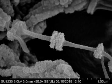
Biomineralized biomaterials
Using biomimetic processes to mineralize different synthetic and natural structures has been a focus of interest in biomaterials research for the last 15-20 years. Recently, the mechanism by which materials scientists have found inspiration in biomimetics for this purpose has shifted from reproducing nucleation and growth of minerals on natural substrates to using amorphous mineral precursors that are controlled by proteins and other biomacromolecules. We have applied these biomimetic methods, such as the polymer-induced liquid precursor (PILP) process or the enzyme-controlled mineralization, to study basic concepts of the mechanism and resulting ultrastructure of bone [PLoS ONE, (2013) 8, e76782], develop surfaces with controlled nanotopographical and mineralized features [Advanced Healthcare Materials, (2014) 3, pp1638], and infiltrate scaffolds made of elastin-like polymers with minerals so that we preserved their original microporosity and intrinsic shape [ACS Applied Materials and Interfaces, (2015) 7, pp25784].
Bioimaging
Virtual Dental Patient (VDP)
The Virtual Dental Patient is a special software program that was developed by Dr. Ralph DeLong and supported by a NIDCR grant (DE09737). The Virtual Dental Patient is an interactive 3D computer rendition of a dental patient that accurately reproduces the surface anatomy of hard and soft dental tissues, tooth contacts, and jaw motion while the teeth are in contact. Because of its numerical nature, the VDP is easily stored, enabling comparisons of sequential VDPs. Quantification of the differences between sequential VDPs provides valuable information for diagnosis, prognosis and outcome assessment of the patient's dental health. The VDP represents a paradigm shift in clinical measurement for dentistry.

The Virtual Dental Patient can be used to go beyond the traditional dental examination. The VDP provides visibility, numerical measurement (including volume and depth changes), and comparison with a previous time period. The VDP provides an environment of near photographic quality, with hard and soft tissue textural details and anatomical relations preserved under static or dynamic conditions. Finally, the VDP enables a dentist to detect subclinical change. Changes below the threshold of chairside observation are now made visible; therefore, adverse incipient dental disease can be detected before it becomes a clinical problem.
Realistic appearance simulation
Dental caries remains one of the major oral diseases. The diagnosis and staging of caries (especially early caries) is a challenging task, given their highly irregular morphologies and appearances. Overdiagnosis often results in unnecessary intrusive restorative procedures. It would therefore be very useful to have a computer simulation tool to train dentists to diagnose and stage caries accurately.
Another area of dentistry where computer graphics can be useful is the computer-aided de- sign/computer-aided manufacture (CAD/CAM) of dental restorations. Although CAD/CAM is now widely used in the production of dental restorations, the focus is mainly on producing the geometry of the restoration precisely. The selection of colors and shades for the restorative materials is still very much a manual process, relying largely on shade guides. Given that the appearance of materials depends greatly on the lighting conditions, a restoration that appears compatible with the neighboring teeth in a dental clinic or laboratory may stand out under different lighting conditions.
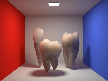
In collaboration with computer scientists, we have been developing photo-realistic rendering techniques for the visualization of translucent objects such as teeth and dental restorations. The figures below show the renditions of some teeth that are synthesized through the simulation of light scattering within the objects [Computer Graphics Forum, (2015) 34, pp585]. The simulation parameters used in the rendering were deduced from the amount of light transmitted and reflected from the sample.


Rendering of teeth: (Left) Three teeth in Cornell box. (Right) Enamel with various translucencies.
Recent Publications
Radfar S, Alikhasi M, Khorshidi S, Tohidkhah S, Morvaridi Farimani R, Shahabi S. Evaluation of One-and Two-Step Impression Techniques and Vertical Marginal Misfit in Fixed Prothesis. International Journal of Dentistry. 2023 Feb 21, 2023.
Shareghi A, Ahmadpoor E, Lori Goiini Z, Ahmadi F, Tohidkhah S. Efficacy of Calcium Hydroxide/Saline versus Calcium Hydroxide/Artemisia persica Essential Oil as Intracanal Medicament to Improve Radiographic Visualization of Periapical Lesions in Necrotic Teeth: A Randomized Clinical Trial. International Journal of Dentistry. 2023 Feb 16, 2023.
Mutreja I, Maalej N, Kaushik A, Kumar D and Raja A.High atomic number nanoparticles to enhance spectral CT imaging aspects, 2023, Materials Advances, 4, 3967-3988.
Kumar D, Mutreja I and Kaushik A. Recent advances in Noble metal nanoparticles for cancer theranostics,. 2023, Journals of Nanotheranostics, 4(2), 150-170.
Mutreja I, Kumar D, Kaushik A and Mishra Y. Manipulative efficient lipids nanostructures for high-performance Dentistry Application, 2023, Journals of Materials Chemistry B, 11, 6159-6160.
Dewey M J., Collins A J.,Tiffany A, Barnhouse V R., Lu C, Kolliopoulos V, Mutreja I, Hickok N J., Harley B A.C., Evaluation of bacterial attachment on mineralized collagen scaffolds and addition of manuka honey to increase mesenchymal stem cell osteogenesis,. 2023, Biomaterials, 294:122015
Kumar D, Moghiseh M, Chitcholtan K, Mutreja I, Lowe C, Kaushik A, Butler A, Sykes P, Anderson N, Raja A LHRH conjugated gold nanoparticles assisted efficient ovarian cancer targeting evaluated via spectral photon-counting CT imaging: A proof-of-concept research, 2023, Journals of Materials Chemistry B, 11, 1916-1928
Ding, H., Z. Cui, E. Maghami, Y. Chen, J.P. Matinlinna, E.H.N. Pow, A.S.L. Fok, M.F. Burrow, W. Wang, J.K.H. Tsoi, Morphology and mechanical performance of dental crown designed by 3D-DCGAN. Dent. Mater., 2023. 39: 320-332.
Chen, Y.C. and A. Fok, Shape optimization of a 2-unit cantilevered posterior resin-bonded fixed dental prosthesis. J. Prosthet. Dent., 2023. 129(1): 180-191.
Tonin, B.S.H., J. Fu, Y. He, N. Ye, H.P. Chew, A. Fok, The effect of abutment material stiffness on the mechanical behavior of dental implant assemblies: A 3D finite element study. J. Mech. Behavior Biomed. Mater., 2023. 142: 105847.
Lin, F., X. Feng, R. Ordinola-Zapata, B. VanHeel, A.S.L. Fok, Load capacity and fracture modes of instrumented tooth roots under axial compression. Dent. Mater., 2023. 39(10): 938-945.
Albelasy, E. H., Chen, R., Fok, A., Montasser, M., Hamama, H. H., Mahmoud, S. H., Abdelrehim, T., & H. P. Chew, (2023). Inhibition of Caries around Restoration by Ion-Releasing Restorative Materials: An In Vitro Optical Coherence Tomography and Micro-Computed Tomography Evaluation. Materials (Basel), 16(16). https://doi.org/10.3390/ma16165558
Sedky, R. A., H. P. Chew, Nour, K. A., Abuelsadat, S. M., Elsherbini, D., & Fok, A. S. L. (2023). Interfacial integrity of bulk-fill resin composite restorations in deep Class-II cavities. Dental Materials Journal
Abdelrehim, T., Salah, M., Conrad, H. J., & H. P. Chew (2023). Auto-Segmentation and Quantification of Non-Cavitated Enamel Caries Imaged with Swept-Source Optical Coherence Tomography. Diagnostics (Basel), 13(23). https://doi.org/10.3390/diagnostics13233586
Kumar, M. S., He, R., Feng, L., Olin, P., H. P. Chew, Jardine, P., Anderson, G. C., & Hong, J. (2023). Particle generation and dispersion from high-speed dental drilling. Clin Oral Investig, 27(9), 5439-5448
Ghani, S. M. A., Hassan, M. I. A., Abdullah, A. H., Ghani, A. R. A., Izra'ai, S. I., Aregawi, W., H. P. Chew, & Fok, A. (2023). Linear and volumetric shrinkage displacements of resin composite restorations with and without debonding. Dent Mater J, 42(5), 659-668.