Polymerization Shrinkage

Polymerization shrinkage of restorative materials remains a major concern because it can cause residual stresses that have been associated with various clinical symptoms. Bi-axial strain gauges are used to determine the post-gel shrinkage component (responsible for residual stress development) during polymerization.
More detailed, full-field shrinkage strain distribution of resin composites, including the depth of cure, can be provided using digital image correlation (DIC).
Fracture Mechanics
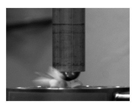
Fatigue Analysis
Because repetitive loading is characteristic for the oral biomechanical environment, dental systems are subjected to fatigue. Failure mechanisms and crack propagation can be studied through the measurement of progressive changes in structural compliance during fatigue loading in a dedicated servo-hydraulic MTS testing system.
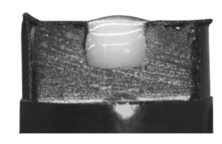
Fracture Toughness

Fracture resistance or toughness is a major requirement of dental hard tissues and their synthetic replacement. It is a major parameter in the development and optimization of new materials for clinical chairside use. The MDRCBB makes extensive use of the small rod methodology, which is appropriate for structures of tooth size dimensions. The methodology gives extensive information about failure mechanisms in brittle and visco-elastic materials.
Water Contact Angels
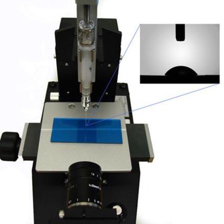
Surface free energy, wettability, lyophilicity and adhesion force are physical properties that can be measured by a goniometer that measures the contact angle of liquids on tested surfaces at the solid/air interface. Measuring the contact angle of a water droplet on a given surface is a quick and easy way to evaluate those properties. The Wilhelmy Balance is an instrument that can also measure the interaction between liquids and solids. It can be used to estimate the energy of solid surfaces. These equipment are used as a tool to analyze the surface properties of newly developed materials.
The oral environment is a challenging electrolyte for metallic materials that are used in the mouth. Orthodontic brackets, bands, and wires, dental implants and partial dentures are made of different metallic materials that can be tested for their resistance to corrosion using potentiodynamic techniques as well as by electrochemical impedance spectroscopy (EIS). Our potentiostat is able to apply a wide variety of electrochemical tests to dental biomaterials susceptible to be corroded in the mouth.
The physical and chemical state of dental biomaterial surfaces is critical for their specific performance in the clinical field. The MDRCBB houses additional equipment to modify (plasma cleaner) and characterize (ATR-FTIR, Attenuated Total reflectance Fourier-Transformed Infrared Spectrometer) specific properties of the materials surfaces.
The mechanical properties of the surface of a biomaterial are critical to predict wear and fatigue performance. Microhardness is a well stablished and very effective method to characterize the mechanical performance of the materials surface in different scales, Vickers and Knoop.
Micro-CT
The center has a versatile X-ray micro-computed tomography (micro-CT) system which can provide high-resolution digital imaging of the microstructures of dental biomaterials and restorations. The system is being used to nondestructively assess the quality of restorative procedures, monitor the development of caries in tissues subjected to different demineralizing and remineralizing regimes, characterize the architecture of scaffolding materials, etc.

Scanning Electron Microscopy
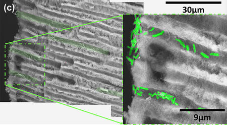
Bacterial Infiltration of Dentinal Tubules
Scanning electron microscopy (SEM) is a useful tool for characterizing morphology of materials. It has been used extensively to evaluate interfacial degradation of dental restorations and the wear properties of dental materials. Careful analysis of the morphology on these materials may show surface breakdown, micro-cracking, voids, localized particle fractures, tensile cracks, and smears on the wear surface, as well as changes occurring on the opposing enamel and on tooth micro-structure. The technique is used for the elemental chemical analysis using X-Ray Energy Dispersive Spectroscopy (EDS). SEM-EDS is used routinely to examine the fracture surfaces of tooth specimens and restorations, the demineralization of tooth tissues, corrosion patterns of metals used in implant dentistry and orthodontics, and topographical and chemical changes of biomaterials surfaces after different manufacturing processes.
Here we image bacterial infiltration of dentinal tubules.